Diagnostics
 Diagnosing Back Pain
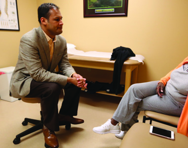 Evaluation of back and neck pain requires a physician
experienced in diagnosing spinal conditions. The work-up begins
with a detailed history and physical examination. Your medical
history helps the doctor understand your back and neck pain and
the influence of your lifestyle on your pain.
Evaluation of back and neck pain requires a physician
experienced in diagnosing spinal conditions. The work-up begins
with a detailed history and physical examination. Your medical
history helps the doctor understand your back and neck pain and
the influence of your lifestyle on your pain.
During your physical exam, your physician will try to pinpoint the source of pain. Simple tests for flexibility and muscle strength may also be conducted. Diagnostic tests may be ordered to confirm the location and source of your pain.
Diagnostics may include:
- X-rays are usually the first step in diagnostic testing methods. X-rays show bones and the spaces between the bones.
- MRI (magnetic resonance imaging) uses a magnetic field and radio waves to generate highly detailed pictures of the inside of your body. Because X-rays only show bones, MRIs are needed to see soft tissues like discs in the spine. These images help your doctor provide a more accurate diagnosis. This type of imaging is very safe.
- CT scan/myelogram - A CT scan is similar to an MRI in that it provides more diagnostic information about the internal structures of the spine. A myelogram is used to diagnose a bulging disc, tumor, or changes in the bones surrounding the spinal cord or nerves. A local anesthetic is injected into your low back to numb the area. A lumbar puncture (spinal tap) is then performed. A dye is injected into the spinal canal to reveal where problems lie.
- Electrodiagnostics - Electrical testing of the nerves and spinal cord may be performed as part of our diagnostic workups. These tests, called Electromyography (EMG) or Somato Sensory Evoked Potentials (SSEP), assist your spine surgeon in understanding how your nerves or spinal cord are affected by your condition.
- Bone Scan - Bone imaging is used to detect infection, malignancy, fractures and arthritis in any part of the skeleton. Bone scans are also used for finding lesions for biopsy or excision.
- Discography - Discography is used to determine the internal structure of your disc. It is performed by using a local anesthetic and injecting a dye into your disc under X-ray guidance. An X-ray and CT scan are performed to view the appearance of the disc composition to determine if its structure is normal or abnormal. In addition to your disc appearance, your doctor will note if you have pain with this injection. The benefit of a discogram is that it enables the spine surgeon to confirm which disc level is really causing your pain. This ensures that surgery will be more successful and reduces the risk of operating on the wrong disc.
- Injections - Pain-relieving injections can relieve back pain and give the physician important information about the problem, as well as provide a bridge therapy.
 Diagnosing the Cause of Knee Pain
Diagnosing knee problems can be tricky, partly because of the subjective nature of pain. Orthopedic surgeons during a physician exam will look for outward signs of injury to the knee, such as sudden swelling and deformity.
They will also interview the patient to determine if the pain symptoms came on suddenly (e.g. traumatic injury or fall) or if the symptoms worsened gradually over time. Other tests involve manipulating the knee joint to determine if there is too much play in the joint or if specific movements worsen pain symptoms.
X-rays are typically taken of the knee from several angles, which can reveal a fracture and signs of arthritis. But because X-rays show only bone and not soft tissue, an MRI may be needed to reveal ligament problems, meniscus tears and other soft tissue issues.
In many cases, the orthopedic surgeon is playing detective to determine what is causing your knee pain. The goal of a visit to the knee doctor is to determine if the knee pain is caused by ligament strain or joint problems like bone-on-bone abrasion from a cartilage tear.
The orthopedic specialist exam typically involves the following steps:
The medical history
The medical history may include your family as well as your personal story. The reason a doctor asks questions about your siblings or parents is to determine whether inherited conditions, such as arthritis, exist within the family. If so, you may be prone to the same problem. You will be asked questions about how and when you first noticed pain in your knee. This part of the exam may seem tedious, but your answers are crucial. For example, if you reveal that your knee pain occurred precisely when you came down from a rebound and you heard a loud pop . . . the doctor will have a high degree of confidence that you probably tore your anterior cruciate ligament.
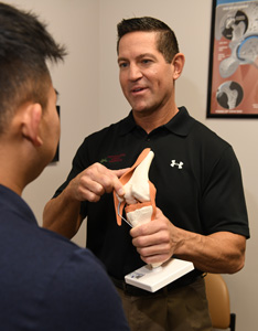
The hands-on knee exam
Each maneuver that the knee specialist performs reveals something only to the doctor who has done thousands of such exams. Some of these knee joint movements may be painful. Some movements of the joint can imply a meniscus tear while other movements can confirm a ligament tear.
X-rays and/or MRI diagnostic tests
Unlike X-rays that only show bones clearly, magnetic resonance imaging (MRI) scans reveal the soft tissues, such as the meniscus and tendons. They go beyond the X-ray in what they demonstrate, but because of cost, are used sparingly.
Unlike X-rays, MRI uses the power of magnetic fields to draw images of the internal structures of the body. MRI is a relatively safe test and, in fact, safer than most tests that use radiation. However, a person undergoing an MRI must remain very still for some time while the computer is creating the image.
 Diagnostic treatments
In a sense, "diagnostic treatments" may sound like contradictory terms. But there are certain "treatments" that a doctor may try. If they succeed, they may confirm a doctor's suspicions about the cause of your pain. This can include the use of oral medications to reduce inflammation, draining fluid, or injections into the knee joint.
Arthroscopic examination
One tool, both diagnostic and surgical, that has dramatically improved knee care is the arthroscope. Years ago, if a surgeon needed to inspect the knee, he or she would have to make a long incision to enter and visually explore the site. Unfortunately, this incision damaged soft tissues and slowed rehabilitation. The arthroscope changed that. Instead of a three-inch incision, knee surgery is now done through small puncture incisions about one to two mm in length.
[Top]